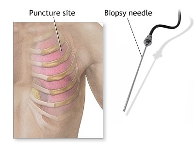
A pleural biopsy is a procedure that removes a sample of tissue from the pleura, the thin membrane that lines the chest cavity and surrounds the lungs. The biopsy is performed to check for disease or infection in the pleura.
-
Needle biopsy
A local anesthetic is injected into the chest, and a special needle is used to extract a tissue sample. Imaging, such as an ultrasound or CT scan, can help guide the needle.
-
Thoracoscopic biopsy
A flexible tube with a light and camera is inserted into the pleural space through a small cut in the chest wall. The tube allows the doctor to see the pleura and take tissue samples.
-
Open biopsy
A surgical incision is made in the chest to access the lung, and a piece of tissue is removed.
A pleural biopsy usually takes 20–40 minutes, and patients are typically able to leave the hospital the same day if there are no complications.
Before the procedure, patients should inform their medical team if they: Have a history of bleeding disorders, Take medications that affect blood clotting, Might be pregnant, and Have any allergies to latex or medicines.
