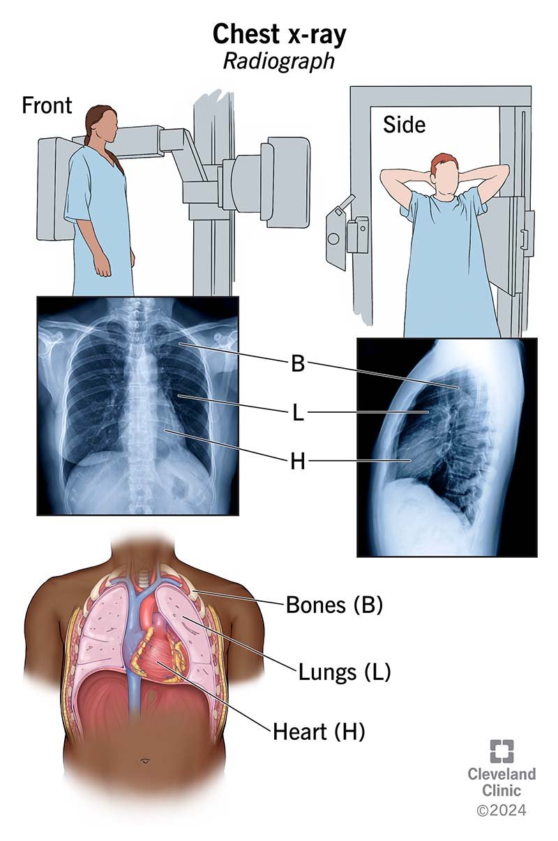
A chest X-ray, also known as a chest radiograph or chest film, is a medical imaging test that uses X-rays to produce a picture of the inside of the chest:
A chest X-ray can help a doctor diagnose conditions affecting the chest, heart, lungs, blood vessels, and other structures:
-
LungsCan detect cancer, infection, air in the lungs, emphysema, cystic fibrosis, and other lung conditions
-
HeartCan show the size, shape, and location of the heart, and may indicate heart failure, fluid around the heart, or heart valve problems
-
Blood vessels
Can reveal aortic aneurysms, other blood vessel problems, or congenital heart disease
During a chest X-ray, an X-ray technician takes a beam of radiation through the chest and records the image on film or a computer. Usually, two images are taken, one from the front and one from the side. The patient is asked to hold their breath when the X-ray is taken.
While there is a slight risk of cancer from the radiation used in a chest X-ray, the benefit of an accurate diagnosis outweighs the risk. Pregnant women should always tell their doctor and x-ray technologist if they are pregnant.
If the results of a chest X-ray are not normal, a doctor may order more specific X-rays or other tests, such as a CT scan, ultrasound, echocardiogram, or MRI scan.
