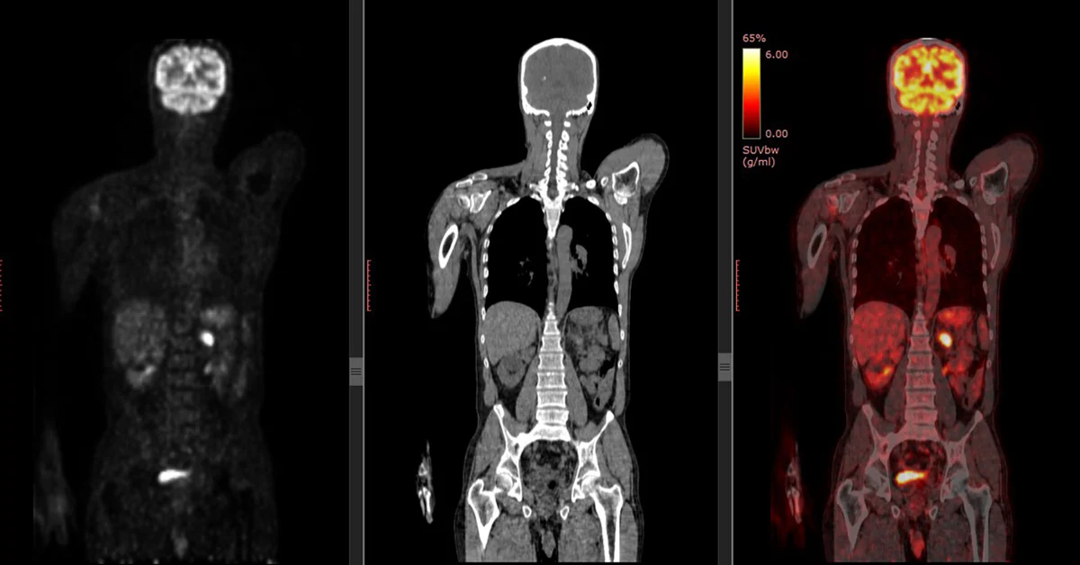
A positron emission tomography (PET) scan is a medical imaging test that uses a radioactive tracer to produce detailed 3D images of the inside of the body. It can show how well organs and tissues are working, and can detect signs of disease, such as cancer, at an early stage.
Here’s how a PET scan works:
- A small amount of a radioactive tracer is injected into the patient’s vein.
- The tracer travels through the blood and collects in areas of the body with high levels of chemical activity.
- The patient lies on a table that slides into a large, doughnut-shaped scanner.
- The scanner detects signals from the tracer and a computer changes them into 3D pictures.
A PET scan is painless, but it can be uncomfortable to lie still for the duration of the scan, which is usually 30 to 60 minutes. The medical team can see and talk to the patient throughout the scan.
PET scans can be used for a variety of purposes, including:
- Diagnosing dementias like Alzheimer’s disease
- Evaluating the brain after trauma
- Detecting cancer and its spread to other parts of the body
- Evaluating the effectiveness of cancer treatment
- Evaluating blood flow to the heart muscle
- Identifying lung lesions or masses
PET scans are often combined with other imaging tests, such as a CT scan or MRI scan, to produce even more detailed images.
